Recent Posts
-

-
 Speaking of HealthBeyond the basics: Exploring advanced breast cancer screening optionsOctober 17, 2023
A closer look at molecular breast imaging: The benefits for dense breast tissue

Women with dense breast tissue have access to a reliable new breast cancer screening called molecular breast imaging (MBI).
This breakthrough technology was developed at Mayo Clinic and is available at Mayo Clinic locations and Mayo Clinic Health System locations in La Crosse and Eau Claire, Wisconsin.
Here are answers to common questions about molecular breast imaging:
How is molecular breast imaging different from a mammogram?
MBI is a fundamentally different approach to breast imaging than mammography.
3D mammography detects slightly more cancers than standard 2D mammography. MBI is significantly better than either type of mammography at detecting breast cancer in women with dense breast tissue.
During a 3D mammogram, individual images are captured by X-ray and combined to simulate "slices" of breast tissue.
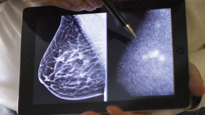
MBI, on the other hand, is a type of functional imaging that uses a special gamma camera to create an image of the breast based on cell activity in the tissue. The images created show differences in the activity of the breast tissue.
During MBI, a small amount of a radioactive tracer is injected into a vein in your arm. The tracer attaches to breast cancer cells that the gamma camera can then detect. MBI identifies tumors based on molecular activity, and areas that absorb more tracer appear as bright spots on the images.
Who should consider molecular breast imaging?
MBI provides the most significant benefit for women with dense breasts. Breast tissue is made up of milk glands, milk ducts, fatty tissue and supportive, or dense, breast tissue. Women with dense breasts have more supportive breast tissue than fatty tissue.
As the density of breast tissue increases, the ability to detect breast cancer with screening mammography becomes more challenging. Both dense breast tissue and cancer appear white on a mammogram, which can make it more challenging to detect whether there is breast cancer in the images.
However, breast density has minimal effect on the detection of breast cancer with MBI screening.
Healthcare professionals may request MBI to help evaluate a breast lump, review an unusual area detected on a mammogram or recommend if other imaging tests produce inconclusive results.
How effective is MBI screening?
Studies show that combining molecular breast imaging and a mammogram leads to finding three times more breast cancers than a mammogram alone.
MBI imaging does a better job of distinguishing between dense tissue, benign tumors and cancerous tissue. MBI also has fewer false positives for breast cancer.
Is molecular breast imaging safe?
Molecular breast imaging is a safe procedure and received Food and Drug Administration clearance over two decades ago in 1999.
The level of radiation exposure during an MBI is comparable to that from a mammogram. It's also less than the natural radiation the body absorbs from the environment annually.
Will molecular breast imaging replace my annual mammogram?
MBI does not replace, but is supplementary to, screening mammography for early breast cancer detection.
For women with dense breasts, the current recommendation is to use 3D mammography and MBI screening together.
What can I expect during a molecular breast image test? 
At the start of the MBI test, you'll receive an injection of the radioactive tracer into a vein in your arm. Fast-growing cells, such as cancer cells, will quickly absorb the tracer.
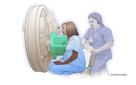
Two small gamma cameras in the molecular breast imaging system detect gamma rays that the tracer emits.
The breast is placed on top of one gamma camera, and the second gamma camera is lowered on top of it just enough to hold it in place during the test.
The gamma cameras record the tracer's activity for 10 minutes, and then the breast is repositioned for a second image. If you are having both breasts imaged, the process is repeated. Two images are created of each breast, and each image takes about 10 minutes.
If you are told you have dense breasts, as 50% of women do, talk with your healthcare team about breast cancer screening options, including 3D mammography and molecular breast imaging.
Next steps:
- Find breast cancer care near you.
- Explore advanced breast cancer screening options.
- Get answers to the top 10 questions about breast cancer.
- Learn when a lump is more than a lump.
Cameron Leitch, M.D., is a radiologist in Eau Claire, Wisconsin.


